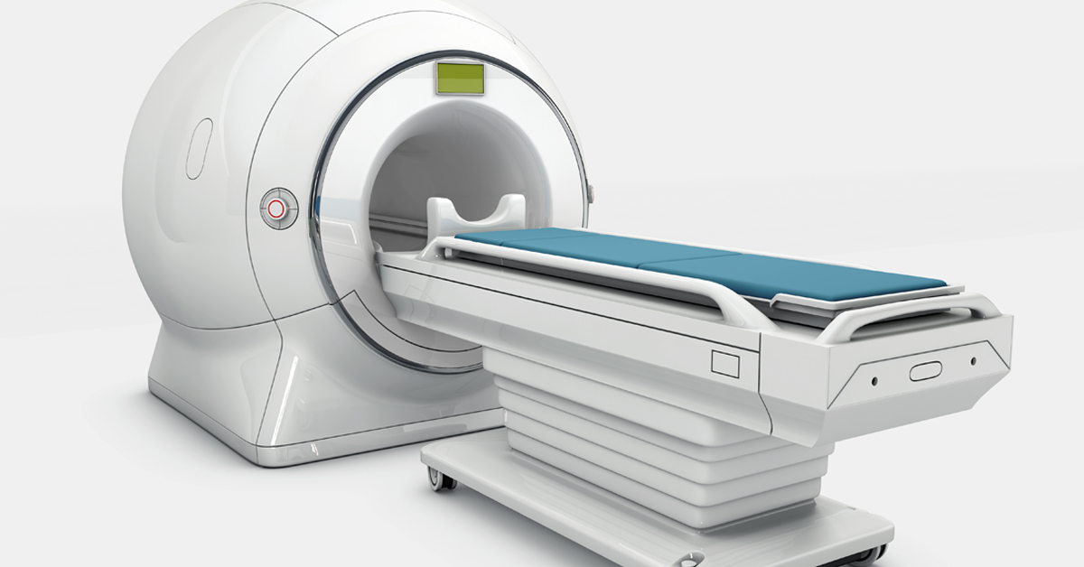
Magnetic Resonance Imaging (MRI): Unlocking the Secrets of the Human Body
In the ever-evolving landscape of medical technology, Magnetic Resonance Imaging (MRI) stands as a shining beacon of innovation. MRI has emerged as a powerful diagnostic tool, offering detailed and non-invasive insights into the human body’s inner workings. This technology has transformed the way medical professionals understand, diagnose, and treat a wide range of conditions.
In this comprehensive blog post, we will embark on a journey through the intriguing world of MRI, delving into its fundamental principles, exploring its applications, and discovering the impact it has on modern healthcare. By the end of this journey, you’ll have a profound understanding of how MRI machines work and why they are a crucial asset in the medical field.
The Science Behind MRI
At the heart of every MRI machine lies the fascinating principles of nuclear magnetic resonance. This science is built upon the behavior of atomic nuclei when exposed to a powerful magnetic field and radio waves. Here’s a simplified breakdown of how MRI works:
Magnetic Field Alignment: When a patient enters the MRI machine, the protons in the hydrogen atoms within their body align themselves with the machine’s strong magnetic field.
Radiofrequency Pulse: A short burst of radiofrequency energy is directed at the patient’s body. This pulse temporarily disrupts the alignment of protons.
Relaxation Process: After the pulse, the protons return to their original alignment, emitting radio waves in the process.
Data Collection: Sensitive antennas in the MRI machine pick up these emitted radio waves, translating them into detailed images.
The data collected during this process allows healthcare professionals to construct highly detailed, cross-sectional images of the patient’s body. These images provide invaluable information for diagnosing a multitude of conditions, from soft tissue injuries to neurological disorders.
MRI vs. CT Scanners: What Sets MRI Apart?
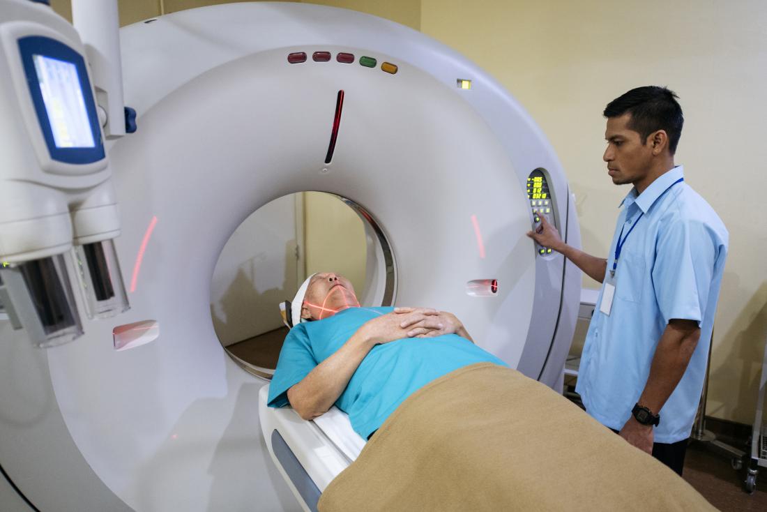
MRI and Computed Tomography (CT) scanners are both critical diagnostic tools, but they work on fundamentally different principles. Understanding the differences between these two technologies is essential for selecting the right tool for specific medical scenarios.
- Radiation Exposure: CT scans involve the use of X-rays, which expose the patient to ionizing radiation. MRI, on the other hand, does not use ionizing radiation, making it a safer option, especially for pregnant women and children.
- Soft Tissue Imaging: MRI excels in capturing highly detailed images of soft tissues, such as the brain, muscles, and organs. CT scans are better at visualizing bones and dense tissues.
- Functional Imaging: MRI allows for functional imaging techniques, like functional MRI (fMRI), which can show brain activity and provide insights into neurological disorders. CT scans do not offer this capability.
- Contrast Agents: Both MRI and CT scans can use contrast agents to enhance image quality. However, MRI contrast agents are generally safer for patients with allergies or kidney issues.
- Specific Diagnostic Applications: Depending on the clinical context, healthcare professionals may choose between MRI and CT scans. MRI is often the preferred choice for neurological, musculoskeletal, and soft tissue assessments, while CT scans are better suited for trauma, bone injuries, and lung assessments.
MRI in Clinical Practice
MRI has found extensive applications in various medical fields. Here are some of the primary areas where MRI shines:
Neuroimaging
MRI has become a cornerstone of neurological diagnostics. It is instrumental in identifying and visualizing brain tumors, strokes, multiple sclerosis, and other neurological disorders. Functional MRI (fMRI) allows researchers to study brain function, aiding in the understanding of cognitive processes and neuropsychological disorders.
Cardiac Imaging
MRI plays a pivotal role in assessing the heart’s structure and function. It can help diagnose heart conditions, such as congenital heart defects, cardiomyopathies, and coronary artery disease. Cardiac MRI provides precise information, enabling physicians to plan effective treatments.
Musculoskeletal Imaging
Orthopedic and sports medicine professionals rely on MRI to assess soft tissues, ligaments, and tendons. It aids in diagnosing injuries like torn ligaments, damaged cartilage, and stress fractures. MRI is essential for creating treatment plans for athletes and individuals with musculoskeletal injuries.
Abdominal and Pelvic Imaging
MRI is invaluable in assessing abdominal and pelvic conditions, such as liver and kidney diseases, tumors, and gynecological issues. Its ability to capture detailed images of soft tissues helps in early detection and accurate diagnosis.
Oncology
In oncology, MRI is used for cancer staging, monitoring tumor response to treatment, and guiding biopsies. Its non-invasive nature and ability to provide 3D images make it a crucial tool in the fight against cancer.
Breast Imaging
Breast MRI is a powerful tool for breast cancer screening and diagnosis. It is particularly useful for assessing high-risk patients and clarifying inconclusive findings from mammography and ultrasound.
The Future of MRI
The field of MRI continues to evolve, promising even more advanced applications and improved patient experiences. Some areas of ongoing research and development include:
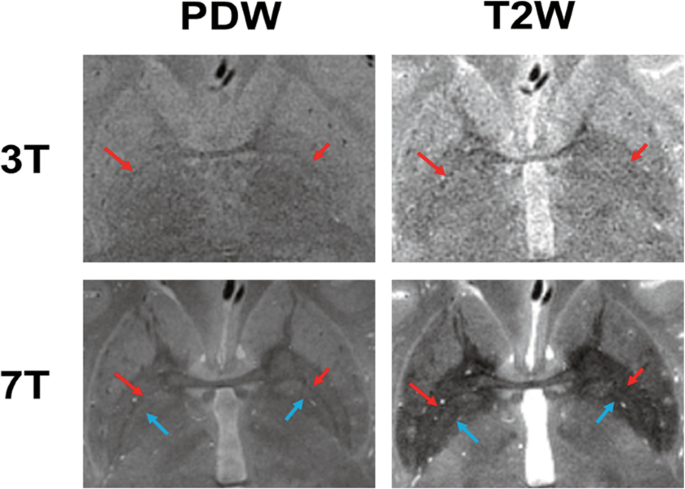
Ultra-High Field MRI: Increasing the magnetic field strength of MRI machines can lead to higher image resolution and shorter scan times. This could significantly improve the diagnosis and monitoring of various medical conditions.
Functional MRI (fMRI): Advancements in fMRI technology are expanding our understanding of the human brain. Researchers are exploring its potential for diagnosing mental health conditions, like depression and anxiety.
Quantitative Imaging: Quantitative MRI techniques aim to provide more than just images; they offer precise measurements of tissue properties. This could lead to earlier disease detection and more personalized treatment plans.
Artificial Intelligence (AI): AI is playing an increasing role in MRI analysis. Machine learning algorithms can assist radiologists by automating repetitive tasks and providing faster, more accurate diagnoses.
Portable MRI: Miniaturized, portable MRI machines are in development, which could bring advanced imaging capabilities to remote or underserved areas.
MRI’s journey from a scientific curiosity to a cornerstone of modern medicine has been nothing short of remarkable. As technology continues to advance, the role of MRI in healthcare will undoubtedly expand, improving patient care, increasing diagnostic accuracy, and enhancing our understanding of the human body.
Conclusion
Magnetic Resonance Imaging (MRI) has transformed the way we diagnose and treat medical conditions. With its non-invasive nature, exceptional soft tissue imaging capabilities, and safety profile, MRI has become an indispensable tool in modern healthcare.
As we move into the future, we can expect even more exciting developments in MRI technology, enabling us to better understand and care for the human body. The journey from discovery to innovation continues, with MRI at the forefront of medical progress. In the world of healthcare, the future is looking exceptionally bright, thanks to the magnetic resonance imaging revolution.


