Introduction:
In the vast landscape of medical technology, one group of machines stands out for its ability to provide intricate details about the human body’s inner workings – Computed Tomography (CT) scanners. These remarkable devices have revolutionized the field of radiology and have become indispensable tools for healthcare professionals worldwide. They offer a level of precision and insight that was once unimaginable. But what exactly are CT scanners, and how do they compare to other imaging technologies, such as Magnetic Resonance Imaging (MRI)? In this blog post, we’ll take an in-depth look at CT scanners, exploring their history, technology, applications, and advancements. We’ll also discuss their relationship with MRI machines, shedding light on how these two vital tools complement each other to enhance patient care.
The Evolution of CT Scanners: From Concept to Reality
To understand the significance of CT scanners in modern medicine, we must first delve into their intriguing history. The concept of computed tomography was initially proposed by British engineer Godfrey Hounsfield in the early 1970s. Hounsfield’s revolutionary idea was to use X-rays from multiple angles to create cross-sectional images of the body, enabling doctors to see inside with unprecedented clarity. His groundbreaking work earned him the Nobel Prize in Physiology or Medicine in 1979.
Over the years, CT technology has undergone substantial developments. The earliest CT scanners were bulky and slow, taking several minutes to generate an image. Today, we have state-of-the-art CT machines that can produce high-resolution images in a matter of seconds. This evolution has not only improved patient comfort but also increased the diagnostic accuracy and expanded the range of applications.
How CT Scanners Work: Unraveling the Technology
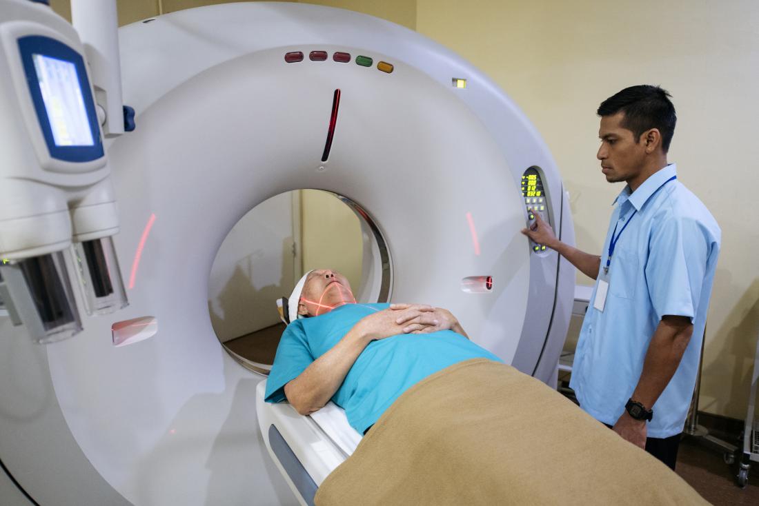
At the heart of every CT scanner is a remarkable piece of technology: the X-ray tube and detector array. These components work together to capture detailed images of the human body. Here’s a simplified breakdown of how CT scanners operate:
X-ray Generation: The X-ray tube emits a controlled beam of X-rays, which passes through the patient’s body.
X-ray Detection: On the opposite side of the patient, a detector array records the X-rays that emerge after passing through the body. The detectors capture the X-rays at various intensities, creating a digital X-ray profile.
Data Reconstruction: A computer processes the data collected by the detectors and uses complex algorithms to create cross-sectional images, or “slices,” of the patient’s anatomy.
3D Image Creation: By stacking these slices together, a 3D image of the scanned area is formed, offering detailed insights into the patient’s internal structures.
Display and Analysis: The final images are displayed on a monitor, where medical professionals can analyze them to diagnose medical conditions accurately.
The ability to create cross-sectional images at various depths within the body is a hallmark of CT scanning. This not only aids in the detection of abnormalities but also provides critical information about their size, shape, and location, which can be vital for surgical planning and treatment.
Applications of CT Scanners: Beyond Diagnosis
CT scanners have found applications in various medical fields, and their utility extends far beyond mere diagnosis. Here are some of the key areas where CT scanners are making a significant impact:
- Trauma and Emergency Care: In cases of traumatic injuries, such as car accidents or falls, CT scans play a pivotal role in quickly identifying and assessing internal injuries. This information guides emergency physicians in making rapid and life-saving decisions.
- Oncology: CT imaging is crucial in cancer diagnosis and staging. It helps in visualizing tumors, determining their size and location, and monitoring the response to treatment. In radiation therapy, CT scans are essential for precise tumor targeting.
- Cardiology: Cardiac CT scans enable the visualization of the heart’s blood vessels and chambers. They are instrumental in diagnosing coronary artery disease, assessing heart function, and planning interventions like angioplasty and stent placement.
- Neurology: CT scans are often used to identify and monitor conditions affecting the brain, such as strokes, intracranial bleeding, and tumors. They help guide neurosurgeons during procedures and assist in post-operative assessments.
- Orthopedics: For musculoskeletal disorders, CT scans provide detailed images of bones and joints. Orthopedic surgeons use this information for accurate preoperative planning and postoperative evaluation.
- Dentistry: Cone beam CT (CBCT) scanners are specialized machines used in dentistry to create 3D images of the oral and maxillofacial regions. These images are invaluable in planning dental implant surgeries and evaluating complex dental conditions.
- Interventional Procedures: CT-guided interventions involve using real-time CT images to guide minimally invasive procedures like biopsies, abscess drainage, and vascular interventions. This precision reduces risks and improves outcomes.
The versatility of CT scanners in various medical specialties underscores their importance in modern healthcare. They provide healthcare professionals with a non-invasive means of obtaining crucial diagnostic information, ultimately leading to more accurate and timely patient care.
Advancements in CT Technology: Making Strides in Patient Care
As with all technology, CT scanners have continued to evolve over the years. These advancements are not only driven by the quest for higher image quality but also by the desire to reduce radiation exposure and enhance patient comfort. Some notable developments in CT technology include:
- Multislice CT: Traditional CT scanners acquired one image slice at a time. Multislice CT, also known as multidetector CT (MDCT), can capture multiple slices simultaneously, significantly speeding up the imaging process. It allows for faster scans, reduces motion artifacts, and provides higher resolution.
- Low-Dose CT: Concerns about radiation exposure have led to the development of low-dose CT techniques. These protocols aim to minimize radiation while maintaining diagnostic quality. They are particularly valuable for pediatric and repeat imaging.
- Dual-Energy CT: Dual-energy CT scanners use two different X-ray energy levels to capture images. This technology offers improved tissue characterization and enhances the detection of certain conditions, such as kidney stones and gout.
- Cone Beam CT (CBCT): In addition to medical applications, CBCT is widely used in dentistry and maxillofacial surgery. It provides high-resolution 3D images with minimal radiation exposure, making it ideal for dental implant planning and orthodontic assessments.
- Artificial Intelligence (AI): AI algorithms are increasingly being integrated into CT scanners to assist radiologists in image interpretation. AI can help identify abnormalities, streamline workflow, and reduce the risk of oversight.
These advancements in CT technology have made imaging more patient-centric, offering quicker results with lower radiation exposure. As a result, healthcare providers can offer more personalized and precise care to their patients, ultimately improving outcomes and safety.
CT vs. MRI: Understanding the Differences
While CT scanners are invaluable tools in the medical field, they are often compared and contrasted with another imaging technology known as Magnetic Resonance Imaging (MRI). Both CT and MRI have their unique strengths and weaknesses, making them suitable for different clinical scenarios.
- Imaging Modalities: CT scanners use X-rays to create images, while MRI machines employ strong magnetic fields and radio waves. As a result, the images they produce are fundamentally different in terms of contrast and information.
- Radiation Exposure: CT scans involve exposure to ionizing radiation, which can be a concern, particularly in repeated or pediatric imaging. In contrast, MRI does not use ionizing radiation, making it safer in this regard.
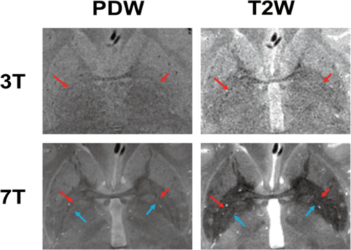
- Soft Tissue Differentiation: MRI is renowned for its superior soft tissue contrast. It excels at distinguishing between various soft tissues, which is particularly useful in neurology, musculoskeletal imaging, and soft tissue tumors.
- Bone Imaging: CT scanners provide excellent bone imaging, making them superior for evaluating fractures, bone tumors, and orthopedic conditions. MRI, on the other hand, may not display bones as clearly.
- Imaging Speed: CT scans are generally faster than MRI, which can be crucial in emergency situations or when the patient has difficulty remaining still.
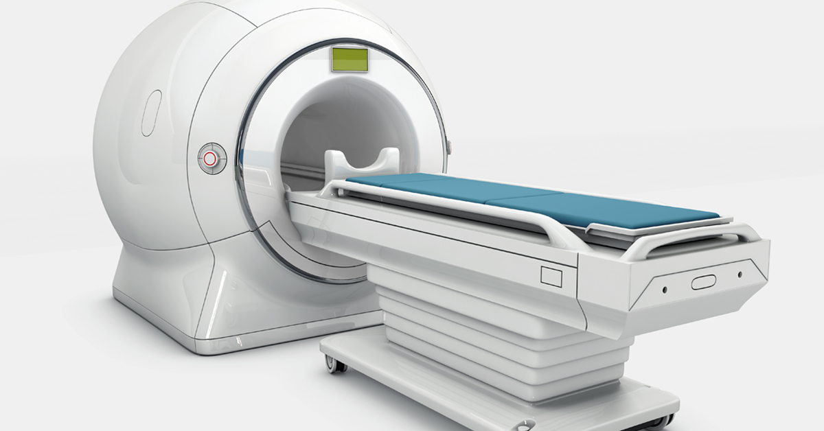
- Claustrophobia: Some patients experience claustrophobia in MRI machines due to their enclosed design, while CT scanners are more open and may be better tolerated by claustrophobic individuals.
- Metal Compatibility: MRI is limited when it comes to patients with metal implants or foreign bodies due to potential safety concerns. CT imaging is generally safe in such cases.
- Functional Imaging: MRI has the edge in functional imaging, such as functional MRI (fMRI) for brain mapping, cardiac stress testing, and diffusion-weighted imaging (DWI) for assessing tissue viability.
In many clinical situations, the choice between CT and MRI depends on the specific diagnostic goals, patient considerations, and the available technology. Medical professionals often collaborate to determine which imaging modality will yield the most accurate and clinically relevant information.
Conclusion: The Endless Potential of CT Scanners
Computed Tomography (CT) scanners have left an indelible mark on the world of medical imaging. Their ability to provide detailed, cross-sectional images of the human body has transformed the way we diagnose and treat a wide range of medical conditions. From trauma care to oncology, cardiology to orthopedics, CT scanners are indispensable tools for healthcare professionals across specialties.
As technology continues to advance, CT scanners have become safer, faster, and more patient-centric. Their integration with artificial intelligence is poised to further revolutionize the field, improving accuracy and efficiency in diagnosis. Moreover, their synergy with other imaging modalities like MRI allows healthcare providers to tailor their approach to each patient’s unique needs.
In the ever-evolving landscape of healthcare, CT scanners remain at the forefront of innovation, serving as beacons of hope and healing for patients worldwide. These remarkable machines will continue to shape the future of medicine, guiding us towards even more precise and personalized care for all.


