Introduction:
Nuclear medicine is a branch of medical imaging that harnesses the power of radioactive materials to diagnose and treat various medical conditions. It is a highly specialized field, and one of its essential tools is the gamma camera. These remarkable devices enable medical professionals to visualize the inner workings of the body at the molecular level, opening up new frontiers in diagnosis and treatment. In this comprehensive guide, we will delve into the world of gamma cameras, understanding their technology, applications, and their critical role in the world of nuclear medicine.
Chapter 1: What Are Gamma Cameras?
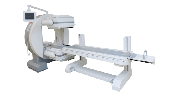
Gamma cameras, also known as scintillation cameras or scintillation detectors, are devices used in nuclear medicine to capture and visualize gamma radiation emitted by radioactive isotopes within the human body. These cameras work on the principle of scintillation, where incoming gamma rays are converted into flashes of visible light. These flashes are then detected, amplified, and converted into digital images that provide valuable information to healthcare professionals.
Chapter 2: The Technology Behind Gamma Cameras
Gamma cameras are intricate pieces of machinery that combine various technologies to produce precise images. They consist of several key components, including:
Crystal Detector: The heart of a gamma camera is a crystal detector, often made of sodium iodide (NaI) or cesium iodide (CsI). When gamma rays interact with this crystal, they produce flashes of light, which are the starting point of the imaging process.
Photomultiplier Tubes (PMTs): These are highly sensitive light detectors that amplify the tiny flashes of light generated by the crystal detector. PMTs are crucial in enhancing the signal and ensuring accurate imaging.
Collimators: These are lead or tungsten devices that help shape and direct the gamma rays towards the crystal detector. Collimators play a significant role in determining the camera’s spatial resolution.
Data Acquisition System: This part of the gamma camera collects and processes the signals generated by the PMTs, converting them into digital images that can be analyzed by medical professionals.
Chapter 3: Applications of Gamma Cameras
Gamma cameras find applications in various fields within nuclear medicine, and their versatility is one of their key strengths. Some of the most common applications include:
Myocardial Perfusion Imaging: Gamma cameras are widely used to assess the blood flow to the heart muscle. This is essential in diagnosing conditions like coronary artery disease.
Bone Scintigraphy: Gamma cameras help detect abnormalities in the bones, such as fractures, infections, and tumors, by tracking the distribution of radioactive tracers.
Thyroid Imaging: In thyroid scans, gamma cameras visualize the distribution of radioactive iodine, helping to diagnose thyroid disorders and diseases.
Single Photon Emission Computed Tomography (SPECT): Gamma cameras are integral to SPECT scans, a specialized imaging technique that provides 3D images of the body’s internal structures.
Sentinel Lymph Node Mapping: In cancer treatment, gamma cameras are used to identify sentinel lymph nodes, which can help determine the spread of cancer within the body.
Chapter 4: Advantages of Gamma Cameras
The use of gamma cameras in nuclear medicine offers numerous advantages, making them indispensable tools in the medical field. Some key benefits include:
Non-Invasive Imaging: Gamma cameras allow for non-invasive imaging, reducing patient discomfort and the risk of infection associated with invasive procedures.
Precise Diagnosis: These cameras provide highly detailed images, aiding in the accurate diagnosis of various medical conditions.
Customizable Imaging: The versatility of gamma cameras enables healthcare professionals to tailor imaging techniques to specific patient needs.
Real-time Imaging: In some cases, gamma cameras can provide real-time imaging, allowing physicians to monitor dynamic processes within the body.
Chapter 5: Challenges and Limitations
While gamma cameras offer many advantages, they also come with their share of challenges and limitations. Some of these include:
Radiation Exposure: The use of radioactive tracers, although minimal, exposes patients to some level of radiation, which must be carefully managed.
Cost: Gamma cameras are complex and expensive devices, making them a significant investment for healthcare institutions.
Resolution Limitations: Achieving high spatial resolution can be challenging, impacting the ability to detect very small abnormalities.
Patient Cooperation: Some imaging procedures may require patients to remain still for extended periods, which can be challenging for some individuals.
Chapter 6: Future Developments in Gamma Camera Technology
The field of nuclear medicine is continually evolving, and gamma camera technology is no exception. Researchers and engineers are constantly working to enhance the capabilities of these devices. Some promising developments include:
Hybrid Imaging: Combining gamma cameras with other imaging modalities, such as positron emission tomography (PET), to provide a more comprehensive view of the body.
Improved Detectors: Advancements in detector materials and technology to enhance sensitivity and spatial resolution.
Reduced Radiation Exposure: Research is ongoing to develop safer radioactive tracers with shorter half-lives, reducing patient radiation exposure.
Artificial Intelligence Integration: The integration of AI algorithms for image analysis, which can help in faster and more accurate diagnosis.
Chapter 7: The Impact of Gamma Cameras on Healthcare
Gamma cameras have had a profound impact on the field of healthcare. They have revolutionized the way diseases are diagnosed and monitored, leading to earlier detection and more targeted treatments. The information provided by gamma cameras helps physicians make critical decisions, ultimately improving patient outcomes and quality of life.
In addition to their diagnostic capabilities, gamma cameras also play a significant role in medical research. They contribute to the advancement of our understanding of diseases and treatment methods, supporting ongoing efforts to find more effective and personalized therapies.
Conclusion:
Gamma cameras are invaluable assets in the world of nuclear medicine. Their ability to provide detailed, non-invasive images of the body’s internal processes has transformed the way medical professionals diagnose and treat diseases. As technology continues to evolve, we can expect gamma cameras to become even more powerful and versatile, further enhancing their role in healthcare. Their journey from a groundbreaking invention to a staple in the medical field is a testament to the relentless pursuit of better healthcare and improved patient outcomes.


